
26 Apr Case Study: MRI Cervical Spine Without Contrast
The Problem
48 y/o right-handed train engineer presents with bilateral hand numbness and right arm pain. He complained of persistent tingling and numbness in his right arm and hand with sudden sharp shock-like pain. This resulted in decreased hand mobility and coordination.
Spinal stenosis usually develops slowly over time. It is most commonly caused by osteoarthritis or “wear-and-tear” changes that naturally occur in our spine as we age. For this reason, the patient was not able to detect any symptoms for a long time, even though some changes might have been seen on his X-rays or other imaging tests he had priorly taken.
He denied neurological symptoms, including clumsiness in the arm, and problems with balance.
Also, loss of function in his hands, like having problems while writing or buttoning his shirt, weakness of the upper or lower extremities, or loss of bowel or bladder continence. The severity of his pain was 10/10.
The Diagnosis
The patient’s medical history was thoroughly reviewed, and a physical examination was conducted, which included feeling his spine and pressing on different areas to see if this caused pain. The patient was also asked to bend in different directions to see if different spine positions bring on pain or other symptoms.
Magnetic resonance imaging (MRI) of the Cervical Spine was done, and it revealed severe stenosis at C4-5, C5-6 & C6-7 with signal change within the spinal cord causing temporary and reversible damage.
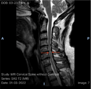
The red arrows in the image demonstrate severe stenosis at C4-5, C5-6 & C6-7.
The Treatment
Choice of stenosis treatments depended on what was causing the symptoms, the location of the problem, and the severity of his symptoms. Because of the complexity of spinal stenosis and the delicate nature of the spine, surgery was considered as all other treatment options had failed the patient.
The patient elected to go for an Anterior Cervical Diskectomy and Fusion with the LDR-C Plate and Cage System from Zimmer/Biomet. What the surgery did was remove the herniated or diseased disc, and a vertebral spacer, bone graft, and metal implants are put in place of the damaged disc and act to fuse the two vertebra together. The overall surgery took only 1 hour and 15 minutes.
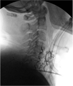
The post-operative x-ray revealed the radio-opaque material, which is the surgical sponge that was placed in the open wound.
The Outcome
48 y/o right-handed train engineer who presented with bilateral hand numbness and right arm pain. The patient went through an Anterior Cervical Diskectomy and Fusion with the LDR-C Plate and Cage System from Zimmer/Biomet. The surgery was performed within 1 hour and 15 minutes only. The patient was discharged the next day without difficulty.
The patient was given pain medications and/or NSAIDs to reduce pain and swelling. He was given a brace to wear for comfort and was encouraged to get up and walk as soon as possible. Our physical therapist recommended a light form of exercise right after spinal surgery to ensure that his back does not stiffen and to reduce swelling. Our physical therapist developed an individualized exercise plan to stretch and strengthen muscles to support his back and stabilize his spine.
He was asked to take hot showers and use hot compresses to help alleviate the pain. The patient will have some swallowing difficulty over the next few days because it was three levels of discs, but he could go back to work as early as one week.